|
|
Knife-Edge Scanning Microscope Brain Atlas
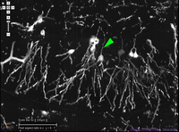
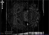
The KESM Brain Atlas (KESMBA) is a web-based, light-weight 3D volume viewer that
serves large volumes (typically the whole brain) of high-resolution mouse brain images (~1.5 TB per brain, ~1 um resolution) from the Knife-Edge Scanning Microscope (KESM), invented by Bruce H. McCormick. [More ...]
[Browse the tutorial]
[Detailed atlas info]
[More info on the atlases ...]
Knife-Edge Scanning Microscope Brain Atlas [Go to the new Atlas]
The enhanced KESM Brain Atlas 2.0 is now available! The new version serves the same data, but it has the following features:
- New data set: Full Golgi data set (Golgi 2) and array tomography data
- Coronal, Sagittal, and Horizontal views: (Golgi and Golgi 2)
- Registration to Allen Brain Atlas (India ink data and Nissl data only)
- Experimental: Array Tomography Data with molecular label overlay.
- SVG-based tiles: faster to load
- Go to the new atlas: http://kesmba.cs.tamu.edu
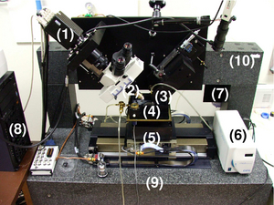
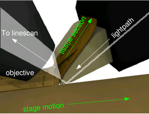
Knife-Edge Scanning Microscope
The data for KESMBA are acquired
using the Knife-Edge Scanning Microscope (Mayerich et al. 2008) shown below. The KESM was invented by Bruce H. McCormick: US patent (2004)
Simultaneous cutting and (line-scan) imaging is the key principle behind the KESM and KESM is the first such instrument to achieve whole mouse brain imaging using the principle Choe et al., Society for Neuroscience Abstract (2009).
 [See the movies]
For more information about the KESM, see the KESM tech spec page.
For more information
Visit the About page.
|
|